
A lead apron is a familiar symbol of radiation protection, but its purpose deserves a closer look. In fluoroscopy rooms, the apron can reduce scatter reaching the torso when staff must remain near the patient. It may also support protection during selected diagnostic procedures. However, it does not replace distance, shielding, exposure-time control, or careful positioning. A practical approach combines all four.
Radiation-protection expert Dr. David Brenner has stated, “There is no threshold below which radiation is completely safe.” This reminder encourages responsible protection, without creating unnecessary fear. A properly fitted lead apron should cover the front of the body, remain closed, and receive regular visual inspections. Creases, cracks, or poor storage can weaken its protective performance. Hang it carefully. Never fold it.
The best choice depends on the procedure, workload, and position of the professional. Some current guidance questions routine apron use when staff stand behind effective structural barriers. Other situations still require personal protective equipment. That distinction matters. A heavy apron can also cause shoulder, back, and neck strain during long procedures. Protection must consider both radiation exposure and physical comfort.
This topic is less simple than it appears. A lead apron is useful, but it is not a complete safety strategy. Reliable protection comes from trained judgment, equipment checks, documented protocols, and continuous review. Even experienced teams can overlook small details, such as an open side panel or an apron stored incorrectly. Good radiation safety remains practical, evidence-based, and willing to improve.
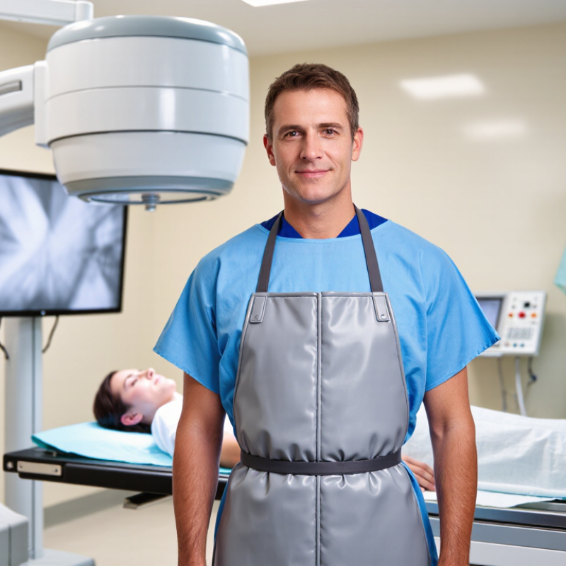
Radiation is energy moving through space or matter. Medical imaging commonly uses ionizing radiation, including X-rays and gamma rays. These forms carry enough energy to change atoms inside living tissue. The effect depends on dose, exposure time, distance, and the body area receiving radiation. Small exposures are carefully managed, but repeated exposure can increase long-term risk.
A lead apron helps reduce scattered X-rays during certain diagnostic procedures. It places a dense protective layer between radiation and sensitive organs, such as the reproductive organs or thyroid. The apron does not block every beam. It also cannot replace distance, short exposure times, or proper shielding. That distinction matters.
In a clinical setting, trained imaging professionals assess whether an apron is appropriate. They check its condition, fit, and position before use. Cracks or folds can weaken its protection. An apron is not a magical barrier. That sounds obvious, yet people sometimes wear it incorrectly or assume thicker always means safer. Protection should follow the procedure, not fear alone. Modern imaging teams also use dose monitoring and the ALARA principle: keeping exposure as low as reasonably achievable while obtaining useful images. I would still question any routine that uses protective equipment without checking current guidance. Good radiation safety depends on informed decisions, careful technique, and honest attention to small details.
During fluoroscopy and radiography, the patient becomes the main source of scattered radiation. Those photons can reach nearby clinicians, assistants, and sometimes patients. A lead apron places an absorbing layer between the body and this scatter. IAEA guidance commonly recommends 0.25–0.5 mm lead-equivalent protection for relevant diagnostic and interventional settings.
The protection is practical, not magical. It mainly shields covered organs, including the torso and reproductive organs. It cannot protect the head, hands, neck, or uncovered back. ICRP Publication 117 emphasizes distance, reduced exposure time, shielding, and good positioning alongside protective clothing.
Standing farther from the patient can dramatically lower dose because scattered intensity follows the inverse-square relationship. Step back when the procedure allows.
Fit and inspection matter. A folded apron can develop cracks. A damaged garment may look acceptable from the outside. Regular fluoroscopic inspection is recommended by professional radiation-safety programs. NCRP Report No. 168 also stresses dose monitoring and equipment controls during fluoroscopically guided procedures.
I have seen staff wear aprons correctly but stand too close to the table. That habit needs reconsideration. Aprons reduce exposure, but they do not replace dosimeters, ceiling-suspended shields, mobile barriers, or trained positioning.
Lead aprons are used when clinical staff may receive scattered radiation during procedures. This commonly occurs during fluoroscopy, cardiac catheterization, pain management, and some orthopedic interventions. They are also useful during mobile X-ray examinations when a person must remain near the patient.
The apron shields the torso from scatter produced when X-rays interact with body tissues. It does not block every exposure. Distance, short exposure time, and fixed barriers remain essential controls. In a busy procedure room, staff may stand beside the table for several minutes. A properly fitted apron can reduce radiation reaching radiosensitive organs, including reproductive organs and bone marrow. A thyroid collar may provide additional protection during longer procedures.
Aprons must stay outside the primary X-ray beam. They should fit comfortably, close fully, and hang flat without folds. Creases can damage the protective material over time. Facilities should inspect them regularly, especially after storage or frequent use. Patients do not automatically need an apron for every diagnostic image. Current practice depends on the examination, positioning, and local radiation-safety guidance. Unnecessary shielding can sometimes interfere with imaging or create a false sense of security.
Small details matter. I have seen teams focus on wearing aprons while standing too close to the radiation source. That habit needs correction. Protection works best as part of a complete safety plan, supported by trained staff and documented clinical protocols.
| Clinical Procedure or Setting | Primary Radiation Concern | When a Lead Apron Is Used | Recommended Protection Approach | Important Limitation |
|---|---|---|---|---|
| Fluoroscopy-guided surgery | Scatter radiation from the patient during prolonged or repeated X-ray imaging. | Routine for staff in the room | Wear a correctly fitted apron, thyroid collar, and other required protective equipment. Maximize distance from the patient and stand on the detector side when practical. | The apron reduces exposure from scatter but does not replace time, distance, shielding, dose monitoring, or equipment optimization. |
| Interventional radiology | Potentially high cumulative scatter exposure during lengthy, high-dose procedures. | Routine for staff in the room | Use wraparound or equivalent protection when movement around the table is required, together with ceiling-suspended shields, table-side curtains, and personal dosimeters. | Lead-equivalent protection is less effective if it is damaged, poorly fitted, or positioned outside the radiation field. |
| Cardiac catheterization and electrophysiology | Repeated fluoroscopy and cine imaging can produce substantial occupational scatter. | Routine for staff in the room | Use an apron and thyroid protection; combine them with ceiling-suspended shields, table skirts, appropriate beam angulation, and dose tracking. | Protective garments should not be folded because creases can damage the shielding material. |
| Mobile C-arm procedures | Scatter from the patient, particularly when personnel remain close to the operating table. | Routine when personnel stay nearby | Wear protective clothing and keep nonessential personnel away. Position staff on the image-receptor side whenever feasible and follow the facility’s mobile C-arm protocol. | The exact dose depends on beam direction, distance, exposure settings, and the time spent near the patient. |
| Routine diagnostic radiography | Short exposures with staff generally able to leave the room or use a fixed barrier. | Usually not needed for staff | Staff should normally stand behind a structural barrier or leave the room. If remaining in the room is necessary, maximize distance and avoid the primary beam. | A lead apron is not a substitute for a controlled area, a protective barrier, or safe positioning. |
| Computed tomography | Scatter from the patient and gantry during image acquisition. | Used only when remaining in the room is necessary | Staff should normally operate from the control room. During exceptional patient-support situations, use appropriate shielding and stand as far from the gantry as practical. | Routine apron use for patients during CT can interfere with automatic exposure control and image quality; follow local radiology policy. |
| Portable radiography at the bedside | Scatter radiation in areas where fixed barriers may not be available. | Used when personnel cannot leave | Nonessential personnel should leave or move as far away as practical. Personnel who must remain should use a protective apron and avoid standing in the primary beam. | Other patients and visitors should be moved away or shielded according to the facility’s radiation-safety procedure. |
| Mammography | Low-scatter diagnostic imaging performed with personnel normally outside the room. | Not routinely required for staff | Staff should use the control-room barrier or leave the room during exposure. Patient shielding decisions should follow current clinical guidance and the examination protocol. | Aprons provide little benefit when the person is already protected by distance or a structural barrier. |
| Dental and panoramic X-ray imaging | Low occupational exposure when the operator uses distance and remains out of the beam. | Not routinely required for operators | Use the recommended operator position or a protective barrier. A patient apron may be considered according to local regulations and current professional guidance. | Never hold the X-ray tube or image receptor during an exposure. |
| Pregnant or potentially pregnant staff | Occupational exposure to scattered X-rays, with additional attention to fetal dose. | Based on duties and exposure assessment | Use the same core controls—time, distance, shielding, and monitoring. Notify the radiation-safety or occupational-health service so duties and monitoring can be reviewed. | Pregnancy declaration does not automatically require removal from radiation work; a lead apron alone does not guarantee fetal protection. |
Why Use a Lead Apron for Radiation Protection?
A lead apron reduces exposure to scattered X-rays during fluoroscopically guided procedures. It does not replace distance, shielding, or personal dosimetry. ICRP Publication 103 sets the occupational effective-dose limit at 20 mSv per year, averaged over five years. NCRP Report 168 also emphasizes controlling staff exposure through time, distance, shielding, and monitoring. Fit matters. The front panel should cover the torso without gaps, while a thyroid collar protects the neck when procedures create significant scatter. Keep it flat.
Inspect the apron before every use. Look for cracks, creases, hardened areas, torn stitching, or uneven surfaces. Never fold it over a chair or leave it on the floor. Store it vertically on a broad hanger, away from heat, moisture, and sharp edges. Do not wash it unless the care instructions allow cleaning. A damaged apron can look acceptable from outside. That is the uncomfortable part. Internal defects may remain invisible.
Routine checks should be documented. Many medical-physics programs use visual inspections at least annually, with radiographic or fluoroscopic testing when damage is suspected. Local policy may require more frequent testing. A qualified radiation-safety professional should assess questionable garments and record their identification, inspection date, findings, and final decision. The U.S. FDA and professional guidance both stress that protective garments require proper maintenance, not indefinite use. Staff should also review posture and positioning. An apron cannot protect areas it does not cover.
A lead apron can reduce exposure from scattered radiation during fluoroscopy, radiography, and other imaging procedures. It is most useful when staff must remain near the patient. The apron does not block every radiation risk. It cannot replace distance, shielding, or careful positioning. It also offers limited protection against the primary beam and may leave the head, neck, arms, and legs exposed. Heavy aprons can cause discomfort, restricted movement, and poor posture. That matters during long procedures. Protection should fit properly and remain free from folds, cracks, or hidden damage. A visual inspection alone may miss defects, so scheduled quality checks are important.
Tips: Stand as far from the source as practical. Step behind a fixed barrier when possible. Use ceiling-mounted screens and table shields correctly. Wear a thyroid collar when appropriate. Keep hands out of the beam. Wear a personal dosimeter according to local procedures. Never place a folded apron on a chair; hang it to prevent damage.
Good radiation safety depends on several controls working together. Time, distance, shielding, equipment maintenance, staff training, and exposure monitoring all matter. The apron may feel reassuring, perhaps too reassuring. A common mistake is treating it as permission to stay closer for longer. That assumption needs regular discussion during team briefings. Even experienced workers can become careless in a busy room. Supervisors and qualified radiation safety professionals should review procedures, exposure records, and workplace habits.
Thank you for reaching out.
Your message has been received, and a member of the Bluestem Energy Solutions team will be in touch soon. We appreciate your interest and look forward to connecting.
